Introduction:
Amniotic band syndrome (ABS) is a rare congenital malformation complex. In the past, gross deformity of the limbs distal to the constricting band and pseudoarthrosis of the affected bones was considered as one of the indications for surgical amputation of the affected limb1. We report a case of ABS in a preterm infant, where simple release of constriction band with z plasty resulted in the growth of mature bone in the tibia and fibula, replacing the pseudoarthrosis and hence did not require surgical amputation.
Case Report
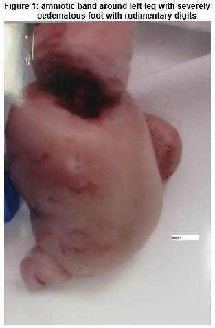
A preterm female infant was born at 29 weeks of gestation with birth weight of 1,384 g to a primi gravida mother. The baby was noted to have constrictive bands around right ring and little fingers, evidence of auto amputation of right and left index and middle fingers with pseudosyndactyly. There were amniotic bands around left lower leg with severely oedematous foot and rudimentary digits (Figure 1). The skin was well perfused and the posterior tibial artery pulsations were easily palpable with no evidence of acute compartment syndrome.
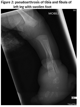
Skeletal survey done on day 3 of life showed constriction of the left lower leg with deficiency of mid to distal tibia and fibula with pseudoarthrosis. The limb deformities were managed with splints, positioning and physiotherapy. She underwent Z-plasty with soft tissue release on the left lower leg on day seven of life.
At 36 weeks of post-menstrual age, a repeat x ray showed markedly improved growth of tibia and fibula with mature new bone formation (Figure 2) and hence she was discharged home.
Discussion
ABS is a rare condition with a prevalence ranging from 1:11,200 to 1:50,5792. The spectrum includes limb deformities and auto-amputations at various levels, pseudosyndactyly, polydactyly and talipes. Facial anomalies, cranial deformations, neural tube defects and other internal organ anomalies have also been well described3,4. Vascular compromise distal to the amniotic band can lead to gangrene of the limb. Hence, careful monitoring and timely release of the constricting bands is essential5-7. Though there was significant swelling of the soft tissue distal to the constricting band, fortunately, our case did not develop any acute vascular compromise. Pseudoarthrosis of the tibia and fibula can cause problems with healing and reunion and might require multiple surgeries. In view of poor outcomes, in the past, early amputation used to be given a strong consideration1. Improved results recently suggest that early amputation may not be necessary, and every attempt should be made to achieve reunion with excision of the pseudoarthrosis, bone grafting and adequate fixation. A newborn infant with ABS of the right leg with pseudoarthrosis of tibia was reported by Rajoo et al. The foot was swollen with a bluish discolouration. The amniotic band which encircled the right leg distally extending deep to the bone was excised urgently and the skin closed with multiple Z–plasties. Postoperatively, the radiograph showed complete remodelling of the tibia with no pseudoarthrosis after six weeks5. Ho et al described the case of a one-day-old neonate born at 28 weeks of gestational age with ABS of forearm with pseudoarthrosis of the radius and ulna. Severe edema occurred in the forearm after fluid resuscitation. This necessitated an urgent operation with release of constriction bands using longitudinal incisions over the forearm. Circulation was restored, and the pseudarthrosis healed with no further surgical intervention 9. Zionts et al described a case of twins with ABS. One twin had pseudoarthrosis of radius and ulna with severe edema, distally. The affected infant’s arm was immobilized with plaster splint, elevated and observed for 10 days. At four months of age, radiographic union was observed and a staged Z-plasty was successful at seven months10.
In summary, our case confirms the potential for bone growth in ABS, once the constricting band is released. Hence, one needs to be cautious while deciding on surgical amputation, even in the presence of pseudoarthrosis, especially in preterm infants. Early limb preserving surgery with release of the constricting band with an intention to salvage the limb appears appropriate.
Conflict of interest:
The authors declare no conflicts of interest.
Ethical Standards:
The study has been performed in accordance with the ethical standards set forth in the 1964 Declaration of Helsinki and its later amendments.
Informed consent:
Informed consent was obtained from the study participants prior to their inclusion in the study. Details that might disclose the identity of the subject under study is omitted.
Correspondence
Dr Anitha Ananthan, MD, Department of Neonatal Paediatrics, Princess Margaret Hospital for children, Roberts Road,Subiaco,WA,6008
Tel: 61893408222, Fax: 61893401266
E-mail: [email protected]
References
1 Lehman WB, Atar D, Feldman DS, Gordon JC, Grant AD. Congenital pseudoarthrosis of the tibia. Journal of pediatric orthopedics Part B. 2000;9:103-7.
2 Froster UG, Baird PA. Amniotic band sequence and limb defects: data from a population-based study. American journal of medical genetics. 1993;46:497-500.
3 Moerman P, Fryns JP, Vandenberghe K, Lauweryns JM. Constrictive amniotic bands, amniotic adhesions, and limb-body wall complex: discrete disruption sequences with pathogenetic overlap. American journal of medical genetics. 1992;42:470-9.
4 Robin NH, Franklin J, Prucka S, Ryan AB, Grant JH. Clefting, amniotic bands, and polydactyly: a distinct phenotype that supports an intrinsic mechanism for amniotic band sequence. American journal of medical genetics Part A. 2005;137a:298-301.
5 Rajoo RD, Mennen U. Congenital constriction band associated with pseudarthrosis and impending gangrene. A case report. South African journal of surgery Suid-Afrikaanse tydskrif vir chirurgie. 1991;29:27-9.
6 Zych GA, Ballard A. Congenital band causing pseudarthrosis and impending gangrene of the leg. A case report with successful treatment. The Journal of bone and joint surgery American volume. 1983;65:410-2.
7 Weinzweig N. Constriction band-induced vascular compromise of the foot: classification and management of the "intermediate" stage of constriction-ring syndrome. Plastic and reconstructive surgery. 1995;96:972-7.
8 Khan T, Joseph B. Controversies in the management of congenital pseudarthrosis of the tibia and fibula. The bone & joint journal. 2013;95-b:1027-34.
9 Ho CA, Richards BS, Ezaki M. Congenital band syndrome with pseudarthrosis of the radius and ulna and impending vascular compromise: a case report. Journal of pediatric orthopedics. 2014;34:e14-8.
10 Zionts LE, Osterkamp JA, Crawford TO, Harvey JP, Jr. Congenital annular bands in identical twins. A case report. The Journal of bone and joint surgery American volume. 1984;66:450-3.
(P570)