Band Heterotopia: An Unusual Cause of Seizures
A Keraliya, P Naphade, D Murphy
Radiology Dept, Brigham & Women’s Hospital, Boston, USA
Introduction
Gray matter heterotopias are characterized by collection of normal neurons located in abnormal locations resulting from interruption of normal neuronal migration from ventricles to cortex. Heterotopias are classified into three groups based on location of abnormal gray matter: Subependymal (periventricular), Subcortical and Band (laminar). Band heterotopia is a rare cortical malformation seen predominantly in females and considered as a mild form of lissencephaly. Here we present the case of a young girl presented with complex seizures, with MRI imaging showing band heterotopia as a cause of her seizures. MRI is particularly useful for establishing the diagnosis of heterotopia and excluding other associated congenital structural anomalies of the brain.
Case report
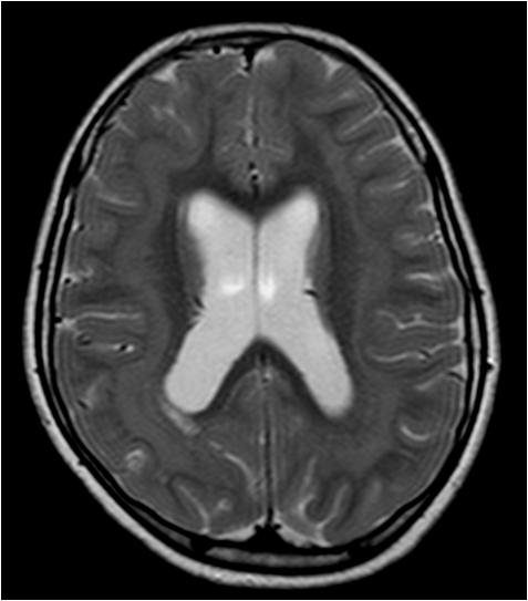
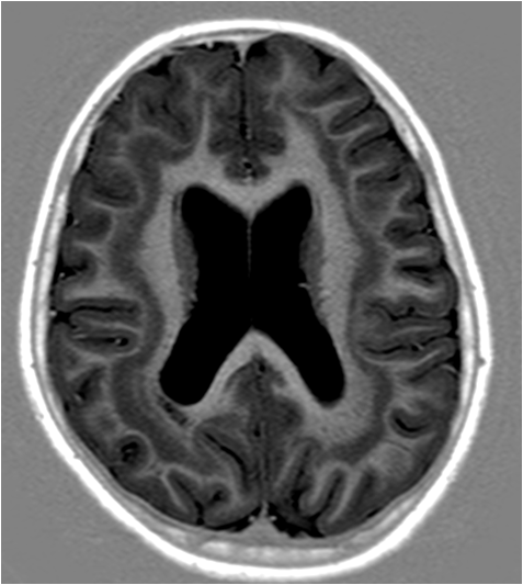
A 12 year old girl presented with four episodes of complex seizures over a period of one year. Clinical examination was unremarkable. The EEG study revealed presence of frequent epileptiform discharges over bilateral parietotemporal regions. MRI brain revealed a band of grey matter located deep to, and roughly paralleling, the cortex and subcortical white matter in both cerebral hemispheres. The abnormal band of grey matter displayed similar signal intensity to cortex on all pulse sequences suggestive of band heterotopia [Figure 1a]. No other structural abnormality was seen on MRI. This case illustrates classic imaging appearance of band heterotopia on MRI. Gray mater heterotopias are characterized by collection of normal neurons located in abnormal locations resulting from interruption of normal neuronal migration from ventricles to cortex. Heterotopias are classified into three groups based on location of abnormal gray matter: Subependymal (periventricular), Subcortical and Band (laminar)1. Partial seizures, developmental delay and mental retardation are common presenting features in patients with heterotopia. Heterotopias are associated other congenital central nervous system anomalies including agenesis of corpus callosum, pachygyria, polymicrogyria, schizencephaly, cephalocele and chiari II malformation.
1a
b
Figure 1a & b: Axial T2 weighted (a) image reveals a laminar T2 hyperintensity located deep and parallel to subcortical white matter. Axial (b) T1 weighted inversion recovery (IR) images show characteristic trilayer appearance with cortex and band of gray matter heterotopia separated by layer of subcortical white matter. The abnormal gray matter is similar in signal intensity to cortical gray matter on both T2 and T1IR weighted images.
Discussion
Band heterotopia is a rare cortical malformation seen predominantly in females and considered as a mild form of lissencephaly. It shows characteristic double cortex appearance on MRI with deep heterotopic gray matter layer parallel to curvature of overlying cortex separated by thin subcortical white matter band. The laminar heterotopic gray matter remains isointese to cortical surface on all sequences and do not enhance after administration of paramagnetic contrast2. MRI is particularly useful for establishing the diagnosis of heterotopia and excluding other associated congenital structural anomalies of the brain.
Correspondence: Dr Abishek Keraliya, Radiology Dept, Brigham & Women’s Hospital, Boston, USA
References
- Abdel Razek AA, KandellAY, Elsorogy LG, Elmongy A, Basett AA. Disorders of cortical formation: MR imaging features.AJNR Am J Neuroradiol. 2009;30: 4-11.
- Barkovich J, Raybaud C. Neuroimaging in disorders ofcortical development. Neuroimaging Clin N Am 2004;14:231–54.
P411


